Protein Yield Quantification Methods in Microplate CFPS Reactions
Choosing the right assay for microplate CFPS requires trading speed, sensitivity.
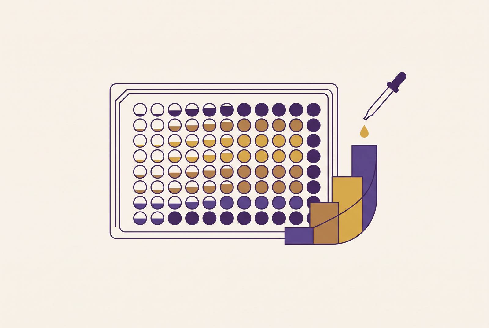
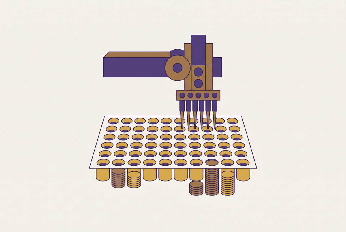
Quantifying protein yield in a microplate CFPS reaction is not a smaller version of quantifying yield in a flask. The volumes shrink, the well counts climb into the hundreds, and the lysate background stays just as messy as it was at bulk scale, so every assay choice has to answer for sensitivity, throughput, and interference all at once. Get the method wrong for the format, and the number on the plate reader tells you about your buffer or your seal, not your protein.
How the microplate format changes the measurement constraints
Volume is the first thing to reckon with. A 384-well low-volume plate can run reactions as small as 5 microliters, and even a 96-well half-area format is at the upper end of what people call "microscale." At 5 microliters, a pipetting error of half a microliter is not noise, it's ten percent of the reaction. Tip wetting alone can eat into that margin, and evaporation over the course of an incubation removes still more. Aluminum seals cut evaporation substantially compared to plastic ones. That is why so many CFPS protocols specify them without much explanation. Skipping that detail means the well in one corner of the plate is not the same reaction as the well in a far corner of the plate by the time anyone reads the plate.
Small volumes also compress the range of signal you can actually detect. Pushing a reaction to the low end of that range causes a sensitive assay to run into its own detection floor; packing too much target into a dense format causes the same assay to saturate. Neither failure looks dramatic on the readout. It just looks like a flat line, and a flat line is easy to misread as "the condition didn't work" instead of "the assay ran out of room."
Throughput adds a second, harder constraint. A screen covering 48 variants, let alone a plate's worth, cannot afford a workflow that requires purifying every well before it can be measured. That rules out entire categories of otherwise good assays right away, because they need a gel, a spin column, or an overnight step that a 384-well screen has no time for. Throughput compatibility is a pass/fail filter applied before anyone even asks how accurate the assay is. It's a pass/fail filter applied before anyone even asks how accurate the assay is.
Then there's the lysate itself. Crude extract carries endogenous fluorescent compounds, metabolic cofactors that absorb light in the same windows most assays use, and total protein that outweighs the expressed target by orders of magnitude. No single assay dodges all of that. Every method on this list trades against lysate background in a different way, and the redesign of prokaryotic CFPS buffers, some formulations have been simplified from 35 components down to 7, shows that the reaction chemistry itself is still being actively rebuilt, and this active rebuilding of the chemistry is what produces the shifting tradeoffs between methods. Whatever you measure has to be calibrated to the specific system generating it, not assumed to transfer from someone else's lysate or someone else's plate.
Finally, batch-to-batch lysate variation means the same reaction run on two different extract preps can produce different absolute signal even when the underlying biology hasn't changed. Normalization, an internal standard, a reference well, RLU expressed relative to a control, has to be built into the plate layout from the start. Adding it after the fact just tells you how much you should have controlled for.
Fluorescent reporter quantification: speed and throughput at the cost of target generality
Superfolder GFP (sfGFP) is the default reporter for a reason: it reads directly on a standard plate reader, no substrate addition, no sample transfer. Instruments like the BioTek Cytation 5 or Synergy H1 read it at 485 nm excitation and 528 nm emission in flat-bottom black half-area 96-well plates, and the whole measurement takes seconds per well.
Raw fluorescence, relative fluorescence units, doesn't mean anything outside the instrument that generated it. Converting RFU into an actual concentration, micrograms or milligrams per milliliter, requires a linear standard curve built from purified sfGFP run on the same plate, same reader, same settings. This gap between reported RFU and real protein mass is a recognized problem in the field, and methods like FPCountR exist specifically to get labs to absolute units without needing to purify a separate standard every time.
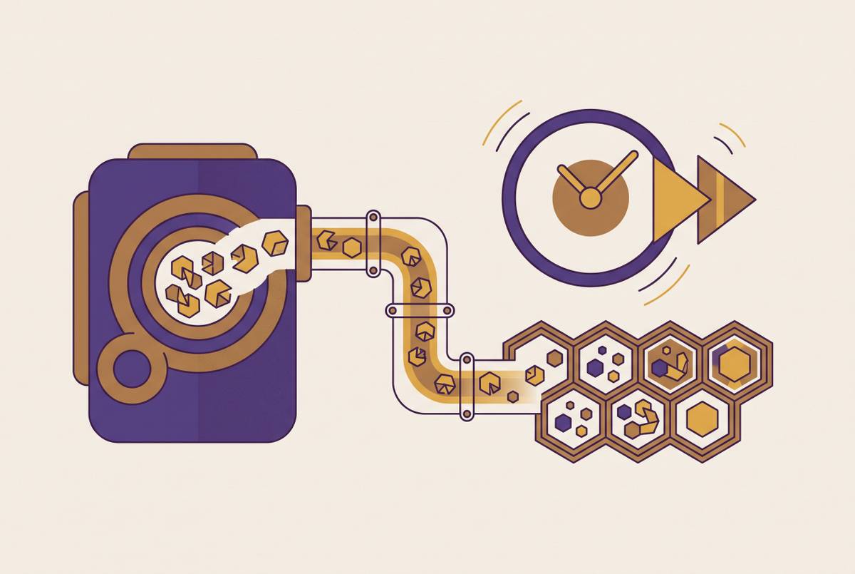
Maturation lag is the more consequential issue, and it's one that catches people who otherwise do everything else right. The GFP chromophore takes time to fold and oxidize after the polypeptide itself is made, and sfGFP is widely used in coupled transcription-translation reactions in part because of its relatively rapid chromophore maturation. Fast is not instant, though. A fixed end-point read taken before the chromophore has fully matured will underestimate total protein made, and that error gets worse, not better, when comparing conditions that change translation speed. A variant that translates fast but matures at the normal rate can look artificially behind a slower-translating variant just because the reading was taken too early.
Split-GFP gets around one obvious limitation: not every protein worth studying is fluorescent. Fusing a small reporter fragment onto the target and co-expressing the complementary larger fragment of that same reporter lets fluorescence form only when the two halves meet inside the reaction, giving a readout for proteins that aren't naturally reporters and largely without disrupting their function. It comes at a cost, though: two templates now compete for the same limited transcription and translation machinery in the reaction, and that competition has to be accounted for when comparing well to well.
None of this gets around the ceiling built into the reporter approach itself. Either the target protein is the reporter, or it tolerates being tagged with one. Plenty of proteins do neither, and fluorescence, regardless of format, never tells you whether the protein folded correctly or does what it's supposed to do. It tells you it exists.
Luciferase reporters: sensitivity advantage and the normalization requirement
NanoLuc (NLuc) solves the background problem that fluorescence never fully escapes. Read with a luciferase assay system on a microplate luminometer, NLuc produces essentially zero background luminescence in a biological matrix, because crude lysate simply doesn't glow on its own. That gives NLuc a signal-to-noise advantage over sfGFP in reactions where endogenous fluorescence or absorbance is already crowding the fluorescence channel.
Running it well means diluting the reaction before reading, since undiluted samples can push the luminometer into saturation, and it means expressing signal as Relative Luciferase Units normalized against a designated control on the same plate, since RLU shifts batch to batch the same way RFU does. Substrate addition, furimazine for NLuc, introduces its own timing question: the signal can vary over the course of a read, so the assay window has to be confirmed for stability before treating one plate's timing as comparable to another's.
The limitations mirror GFP's. NLuc tells you about NLuc, or about a fusion partner tagged with it, not about an arbitrary untagged protein. And RLU normalization corrects for batch drift, it doesn't hand you an absolute mass number. Getting to micrograms per milliliter still needs a purified standard curve or an equivalent calibration step layered on top.
Luciferase earns its place over fluorescence specifically when lysate background is unacceptably high for GFP, or when the reaction runs on a timescale short enough that GFP's maturation lag eats into a meaningful fraction of the total assay window.
Colorimetric activity assays and ELISA: measuring what the protein does rather than how much is present
Enzymes can be measured by what they do, and that changes the question being asked. A colorimetric activity assay measures functional, correctly folded enzyme specifically. For a protease, a kinase, or a biosynthetic enzyme, that's usually the number that actually matters, since a total-protein count can't tell folded protein apart from a well full of misfolded aggregate.
The tradeoff sits in what activity leaves out. A variant that folds poorly but happens to be highly active per molecule can look indistinguishable from one that folds well but turns over substrate more slowly. Activity is a proxy for function.
ELISA plays a similar role for antibody fragments and other binding proteins, where folding quality is the whole point of the screen. The CFPS reaction gets diluted, often around 1:200, and transferred to a 384-well Maxisorp plate coated with the target antigen, then detected with an HRP-conjugated secondary antibody. That measures the antigen-binding-competent fraction specifically, which is exactly the number a nanobody or antibody fragment screen needs, since plenty of protein can be synthesized without any of it binding its target. It's an extra plate-based step, but the 384-well format keeps it compatible with automation and reasonable well counts. What it doesn't give is total protein mass; that requires pairing the ELISA signal with a separate total-expression measurement to back out what fraction of the protein made actually folded correctly.
Bulk-protein assays (BCA, Bradford) and radioactive incorporation: why they do not translate cleanly to microplate CFPS
BCA measures total protein in a sample, and in a crude CFPS reaction, total protein is overwhelmingly lysate protein that was already there before the reaction started. The newly synthesized target is a rounding error by comparison. BCA becomes genuinely useful only after purification, where it can quantify an isolated target like sfGFP against a known standard curve (roughly 0.042 to 0.68 mg/mL in HEPES buffer is a typical working range). Run directly on a crude microplate reaction, though, it mostly reports on how much lysate protein is in the well, not how much product got made.
Bradford has the same structural problem, plus the added complication that certain components of crude lysate preparations can interfere with the Bradford dye-binding chemistry, adding another source of error on top of the background issue it already shares with BCA.
Radioactive amino acid incorporation, using carbon-14 leucine or sulfur-35 methionine, is the most target-agnostic method on this list. TCA precipitation followed by liquid scintillation counting measures new protein synthesis directly, without needing a reporter, a tag, or a folded, active protein. It measures new protein synthesis regardless of what the protein does or whether it fluoresces. What it does require is radioisotope handling infrastructure, waste disposal for radioactive material, and a workflow entirely outside the standard plate-reader pipeline. None of that scales to 384 wells. It belongs in extract characterization and validation work, confirming that a new lysate batch actually supports synthesis at the expected rate.
Western blotting fills a different niche: confirming that the band you're calling "product" is actually full-length. An anti-tag primary antibody, an anti-FLAG tag is a common choice, with chemiluminescent detection gives target-specific, size-resolved confirmation of the expressed product that no plate-reader assay can. It's inherently low-throughput and can't be run per well at microplate scale, which makes it a confirmatory step for a short list of hits coming out of a screen, not a quantification method for the screen itself.
LC-MS for metabolite and absolute yield characterization: depth over throughput
A single LC-MS run can separate and quantify on the order of 40 compounds involved in CFPS metabolism at once: amino acids, nucleotides, and energy intermediates like a key energy-carrying molecule and its breakdown products. That gives a systems-level view of the reaction that no endpoint assay, fluorescent, luminescent, or colorimetric, can offer, because those assays report only the output, not what happened to the inputs along the way.
That distinction matters mechanically. LC-MS can identify whether a reaction ran out of a specific amino acid, lost energy regeneration capacity partway through, or suffered degradation of the DNA template itself before transcription finished, three failure modes that would all look identical as "low yield" on a fluorescence plot. Work on 384-well CFPS optimization posted to bioRxiv identified DNA template stability and amino acid preservation as primary factors sustaining CFPS activity over the course of a reaction, a finding that required metabolic-level measurement to establish. No amount of endpoint fluorescence data would have surfaced it.
LC-MS is not a screening tool, and it was never meant to be one. Sample prep, chromatographic separation, and instrument time per run rule it out for anything approaching plate-scale throughput. Its place is downstream: applied to a handful of representative conditions once plate-based screening has already narrowed a large variant space down to the ones worth understanding mechanistically.
Tradeoff map: matching method to experimental goal in microplate CFPS
For primary screening across a full plate or substantial fraction of one, sfGFP or NLuc reporters are the only realistic options: read in place, no sample transfer, and a standard curve built into every plate run. Whichever one is chosen, the timing of reporter behavior has to be planned for in advance, maturation lag for GFP, substrate addition and stability for NLuc, and an activity assay should replace or supplement the reporter whenever the actual screening goal is function rather than raw expression level.
For antibody or binding-protein campaigns, ELISA run on diluted CFPS reaction output measures the antigen-competent fraction specifically, and pairing it with a co-expressed reporter separates folding efficiency from raw translation level, two numbers that a single assay can't distinguish on its own.
For validating hits after a screen has already narrowed the field, Western blotting with an anti-tag antibody confirms full-length product with a level of specificity no plate-based assay reaches, and BCA or Bradford becomes usable again once the hit has been purified and total protein no longer interferes with the readings.
For extract characterization, lot-to-lot quality control, and yield benchmarking work that sits outside routine screening, radioactive incorporation and LC-MS both belong in the toolkit because they answer questions, true target-agnostic synthesis rate, metabolic bottleneck identification, that no plate-reader assay was ever built to answer.
Sources
- An AI-driven workflow for the accelerated optimization of cell-free protein synthesis
- A simplified and highly efficient cell-free protein synthesis system for prokaryotes
- Quantification of Interlaboratory Cell-Free Protein Synthesis Variability | ACS Synthetic Biology | ACS Publications
- biorxiv.org
- promegaconnections.com
- news-medical.net
- citeqbiologics.com
- jove.com