Membrane Protein Expression Failures in Cell-Based Systems
Researchers can now diagnose why membrane proteins fail to express and know when to abandon cells.

Membrane protein expression failures in cell-based systems trace back to four distinct, well-characterized biological mechanisms, not to bad luck or poor technique. Understanding where each failure comes from gives researchers a way to diagnose what actually went wrong, and a clear basis for deciding when it is time to leave cell-based expression behind.
The structural features that make membrane proteins hard to express
Membrane proteins are built differently from soluble proteins, and that difference is the root of almost every expression problem that follows. Their transmembrane domains are long hydrophobic stretches that need shielding from water at every step, during synthesis, during transport to the membrane, and during insertion. On top of all this, most membrane proteins occur at very low levels in their native organisms, so they were never selected for the kind of high-volume production that overexpression systems are built around.
The four failure modes of cell-based expression and the biology behind each one
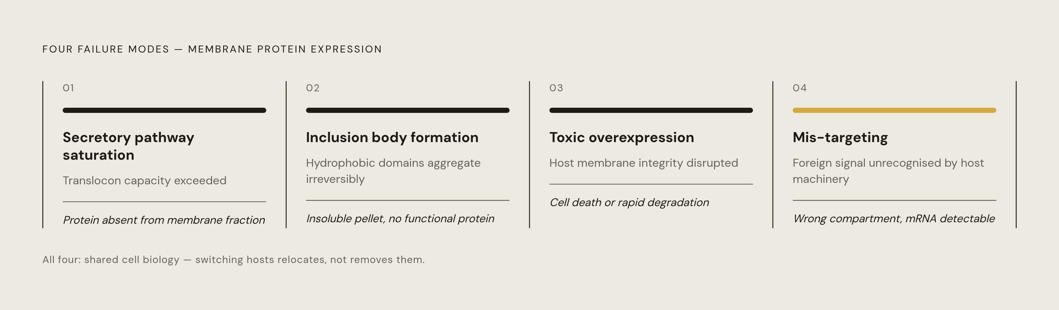
Each structural liability described above maps onto a specific, well-documented failure mode in cell-based expression. None of these four problems are quirks of a particular lab protocol. They are consequences of how cells are built. This same set of mechanisms shows up across organisms and across expression strategies.
Secretory pathway saturation. Membrane proteins reach their destination through the secretory pathway, using the signal recognition particle and the translocon, and that machinery has a fixed capacity. Overexpressing a target floods this pathway past what it can handle, and the cell has no way to process the resulting backlog. The same bottleneck occurs in eukaryotic hosts: the ER translocon is a shared resource, used by every membrane-destined protein the cell needs for its own survival, so overloading it with a foreign target competes directly with the cell's own housekeeping.
Inclusion body formation. When hydrophobic domains miss the translocon, or arrive faster than the translocon can insert them, they aggregate into inclusion bodies instead. These are not simply dormant stockpiles of otherwise-good protein. Inclusion bodies represent a misfolded state that is often irreversible, and attempts to refold protein recovered from them tend to produce low yields and non-native conformations. Reviews of Lactococcus lactis as an expression host point to inclusion body formation, endotoxin production, residual proteases, and overly fast translation rates as the main reasons E. coli so often produces disappointing yields of functional membrane protein.
Toxic overexpression. Many membrane proteins disrupt the host's own membrane once they accumulate past a certain point: ion channels leak, transporters stop working correctly, receptors fire signals the cell never asked for. When a cell faces this kind of disruption, it shuts down expression, degrades the protein it just made, or dies outright, and all three responses destroy yield. Cell-free protein synthesis itself was first developed to study the basic mechanics of protein synthesis and to help decipher the genetic code. Its usefulness for proteins that are toxic to living hosts, recognized only later, was a byproduct of its original purpose.
Mis-targeting and failed membrane integration. A membrane protein introduced into a foreign host may go unrecognized by that host's targeting machinery, or it may be recognized and then routed to the wrong membrane. A protein stuck in the cytoplasm, or sitting in the wrong compartment, counts as a failed experiment regardless of how much of it the cell managed to synthesize. Reviews of cell-free systems for transmembrane proteins confirm that mis-targeting is a documented failure mode even when a foreign host's targeting machinery is structurally similar to the protein's native pathway.
Host-switching relocates the problem rather than solving it
The usual response to a failed membrane protein expression run is to try a different host: move from E. coli to yeast, to insect cells, or to mammalian cells. That move changes the organism, but it does not remove the four failure modes, because those failure modes come from properties that all living cells share. Mammalian cells add cost and complexity without guaranteeing correct topology for proteins that did not evolve in a mammalian membrane. In practice, this uncertainty pushes labs into engineering new strains, co-expressing chaperones, tuning induction conditions, and screening promoters, layering workaround on top of workaround within a system that still cannot separate protein synthesis from the cell's need to stay alive. But the same study found that cells accumulate elevated mRNA levels for high-expressing variants, with the authors suggesting this comes from efficient translation-coupled membrane insertion reducing transcript degradation. That finding confirms expression success in living cells stays tied to the cell's own membrane insertion machinery, no matter which host or how much data is used to predict it.
The cumulative cost of these failures, as revealed by the gap in the Protein Data Bank
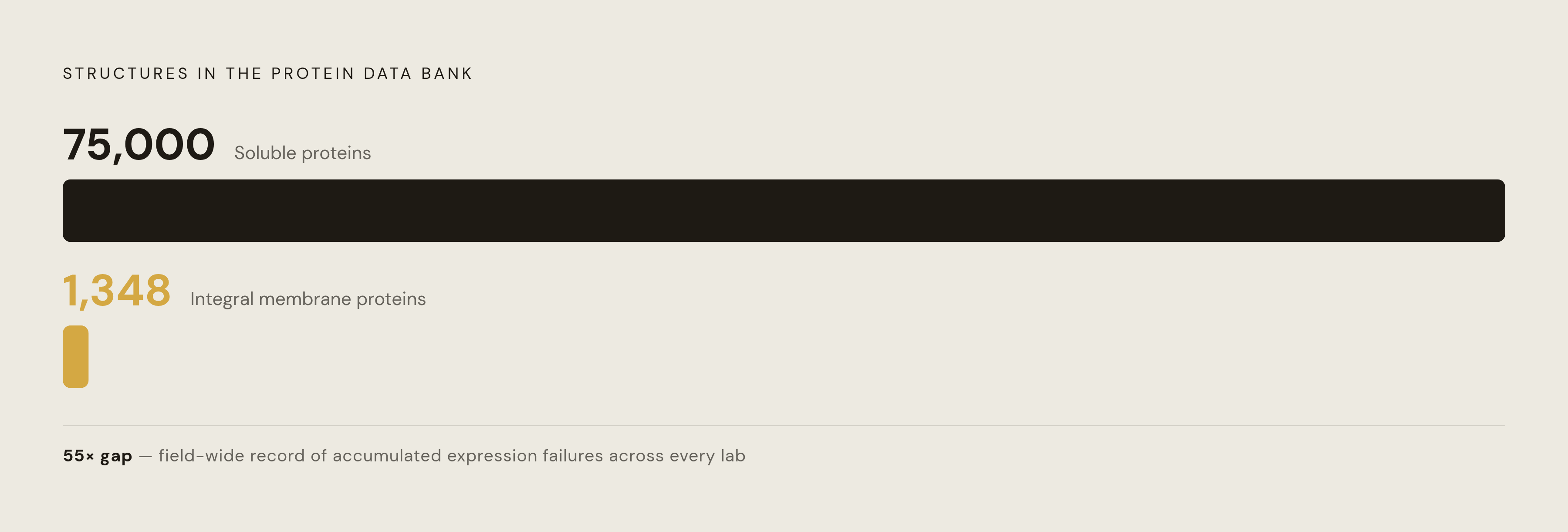
The Protein Data Bank offers a field-wide record of how badly these failures add up. Integral membrane proteins are heavily represented among drug targets, yet they stay underrepresented in structural databases relative to soluble proteins, and that mismatch is the clearest empirical sign of a systemic expression problem, not just a handful of unlucky targets. Over a seven-year period, the number of unique 3D membrane protein structures grew from roughly 400 to 1,348, a figure still dwarfed by the approximately 75,000 structures available for soluble proteins. Reviews of membrane protein expression trace this shortfall directly to the difficulty of getting enough functionally folded protein to work with, so expression failure, not any limitation in structural analysis techniques, is the actual bottleneck. A Nature Scientific Reports study reports that FGFR3-TACC3 failed to express to sufficient quality or functionality in traditional expression systems, and that no published high-resolution structure of any full-length receptor tyrosine kinase exists. That absence follows directly from cell-based expression's inability to handle the combination of high flexibility, a strong tendency to aggregate, and the lack of any established method for extracting the protein within its native lipid environment.
Removing the constraints behind cell-based failures
Cell-free protein synthesis does not just patch around the four failure modes described above. It removes the cellular context that made those failure modes unavoidable. There is no secretory pathway to saturate in a cell-free reaction, because there is no translocon throughput limit at all: synthesis and membrane insertion happen in a defined reaction mixture, at a rate the investigator sets rather than one the cell imposes. There is no viability constraint either, since a toxic protein can be synthesized without anything needing to survive the process. A cell-free extract does not need to stay alive after making the protein it was built to make. Mis-targeting disappears as a concern too, because there is no cellular routing machinery left to confuse: membrane-mimetic scaffolds go directly into the reaction, and the protein folds into them as it comes off the ribosome. Inclusion body formation drops sharply when the right membrane mimetic is present, because the hydrophobic domains have somewhere compatible to fold into the moment they emerge. The open format of a cell-free reaction also lets the investigator adjust redox potential, pH, ionic strength, tRNA composition, and lipid identity directly, adjustments that would compromise a living cell's growth if attempted inside it.
The role of membrane mimetics in getting membrane proteins to fold correctly in cell-free reactions
Removing the cell does not remove the hydrophobic domain's basic need for a membrane-like environment to fold into. Without some kind of scaffold, a transmembrane domain synthesized into an aqueous cell-free reaction aggregates just as it would in a cell's cytoplasm. It simply needs a different solution. Three classes of membrane mimetics have been used to supply that solution: detergent micelles, bicelles, and lipid-protein nanodiscs, and their performance varies substantially. Reviews of cell-free systems for transmembrane proteins report that nanodiscs significantly improve synthesis of integral membrane proteins in soluble, folded form, regardless of which lipid is used, and that nanodiscs produced bacteriorhodopsin at roughly 80% activity while also supporting correct folding of TM-ErbB3, a result detergents could not match. That evidence has made lipid-protein nanodiscs the leading approach for producing folded integral membrane proteins in cell-free systems. A 2025 study in Nature Communications introduced DeFrND, a detergent-free reconstitution method that uses engineered membrane scaffold peptides, chemically modified Apolipoprotein-A1 mimetic peptides, to build native nanodiscs efficiently enough for single-particle cryo-EM work while keeping native lipids intact. That method directly addresses a weakness in traditional nanodisc reconstitution, which normally requires detergent solubilization first, a step that compromises the protein's physiological structure and disrupts natural membrane asymmetry. You can vary nanodisc composition systematically within a cell-free reaction, so lipid identity, scaffold protein, and disc size all become tunable parameters, and you can optimize folding conditions for a specific target instead of accepting whatever the host cell happens to offer.
Two recent cases where cell-free expression succeeded where cell-based systems had failed
Two documented cases show these mechanistic advantages translating into results on targets that cell-based systems could not touch. FGFR3-TACC3 is an oncogenic fusion protein combining fibroblast growth factor receptor 3 with transforming acidic coiled-coil-containing protein 3, and it is clinically relevant across multiple cancers. Using a cell-free system, the ALiCE platform produced functional FGFR3-TACC3 in under 48 hours at 300 micrograms per milliliter of lysate, with demonstrated phosphorylation of the kinase domain, and the protein was reconstituted using SMA copolymers. The second case involves the adenosine A2A receptor, a GPCR. A thermostabilized version of the human receptor, crystallized in lipid cubic phase, was used to screen 568 fragments and identify 23 initial hits, marking the first large-scale fragment screening effort against crystals of a membrane protein grown in lipid cubic phase. That screen depended on obtaining a structurally homogeneous, functionally active receptor preparation, exactly the kind of material cell-based expression routinely fails to deliver for GPCRs.
Using cell-free synthesis to screen membrane protein variants at scale (a workflow that cell-based systems make impractical)
The advantages documented above extend past rescuing single difficult targets. They change what kind of research is practical: you can start cell-free protein synthesis directly from PCR products, skipping cloning and transformation, and you can run multiple variants as parallel reactions in standard plate formats. In April 2025, Nuclera added a membrane-protein workflow to its eProtein Discovery platform, combining cell-free synthesis with digital microfluidics to express, purify, and stabilize functional membrane proteins within 48 hours, a scale cell-based expression cannot reach. In September 2025, LenioBio and AffinityAI combined AI-guided protein design with high-throughput cell-free expression, linking computational design pipelines directly to wet-lab validation. One documented biofoundry workflow expressed a library of 1,217 enzyme variants using cell-free synthesis and screened nearly 11,000 reactions, feeding the results into a machine learning model that predicted high-performing variants as part of a fully automated pipeline covering DNA assembly, expression, screening, modeling, and redesign. The Edinburgh and Bristol preprint mentioned earlier is built on a library of 12,248 computationally designed membrane protein variants, and it is only possible because expression did not need a full cell-based workflow for each individual variant. For membrane protein screening specifically, this throughput advantage compounds in a second way: every variant that would have triggered cell death or secretory pathway saturation in a cell-based system still gets expressed in a cell-free reaction, so the resulting dataset is more complete and carries less bias toward sequences that merely happened to be non-toxic. Platforms built for this purpose, such as Sepia Biosciences' OpenCFPS™, are designed to support milligram-scale production for assays and characterization alongside high-throughput variant screening, with pricing meant to make large-scale cell-free screens accessible beyond the small number of labs with the biggest budgets.
A diagnostic framework for deciding when a membrane protein failure calls for moving to cell-free
Each of the four failure modes leaves a signature a researcher can read to identify an expression failure. Each leaves a distinct signature. Secretory pathway saturation shows synthesized protein, detectable mRNA, ribosomes actively engaged, that never accumulates in the membrane fraction, paired with impaired cell growth right after induction. Inclusion body formation is visible in the pellet fraction after lysis: SDS-PAGE confirms the correct molecular weight, but activity is absent, and refolding attempts recover little or no functional protein. Toxic overexpression shows an inverse relationship between induction level and yield, where lower induction produces healthier cultures but less protein, and growth curves show arrest or death once induction begins. Mis-targeting shows synthesized, soluble protein that fails to cofractionate with the expected membrane, with topology assays revealing an incorrect orientation.
Some situations point clearly toward cell-free expression. A protein that proves cytotoxic in every host tested calls for a system that removes the viability constraint. A protein that depends on a specific lipid environment no available host can supply calls for a system where lipid composition can be set directly. If a project screens dozens or thousands of sequence variants, it calls for the throughput cell-free synthesis makes possible and cell-based systems make impractical. A target that is a receptor tyrosine kinase, a GPCR, or a multi-pass transporter with no existing structural data fits the pattern the FGFR3-TACC3 and β2AR cases already demonstrate.
Other situations still favor working within cell-based expression first. A protein that closely resembles a homolog known to express well in the intended host, where the failure looks like a codon usage or promoter problem rather than a structural one, is worth optimizing before switching platforms. Proteins that depend on post-translational modifications only eukaryotic cellular machinery reliably provides remain better suited to cell-based hosts for now, though eukaryotic cell-free lysates continue to close that gap.
The broader lesson holds regardless of which path a given protein takes: membrane protein expression failures follow known mechanisms, and a researcher who can name the mechanism behind a failure is positioned to choose the right tool. Cell-free protein synthesis succeeds for this class of protein because it removes the mechanisms that cause the failure, not because it treats the symptoms those mechanisms produce.
Sources
- Cell-free systems for expression of transmembrane protein - PubMed
- Effective sequence-to-expression prediction for membrane proteins using machine learning and computational protein design
- Cell-free expression and SMA copolymer encapsulation of a functional receptor tyrosine kinase disease variant, FGFR3-TACC3
- Lactococcus lactis, an Attractive Cell Factory for the Expression of Functional Membrane Proteins